Empowering BioPharma
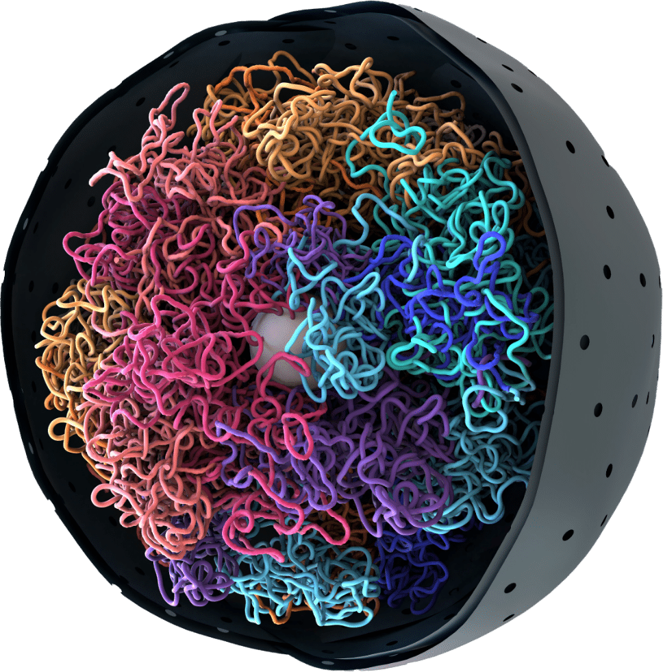
Unravel disease mechanism, uncover drug modes of action, and tap into a rich array of biomarkers. An under recognized component of the epigentic landscape, 3D genomics bridges the intricate gap between genotype and phenotype. This transformative technology propels research and development, empowering biotech and pharmaceutical companies with unprecedented insights to revolutionize therapeutic strategies and advance precision medicine.
Learn More